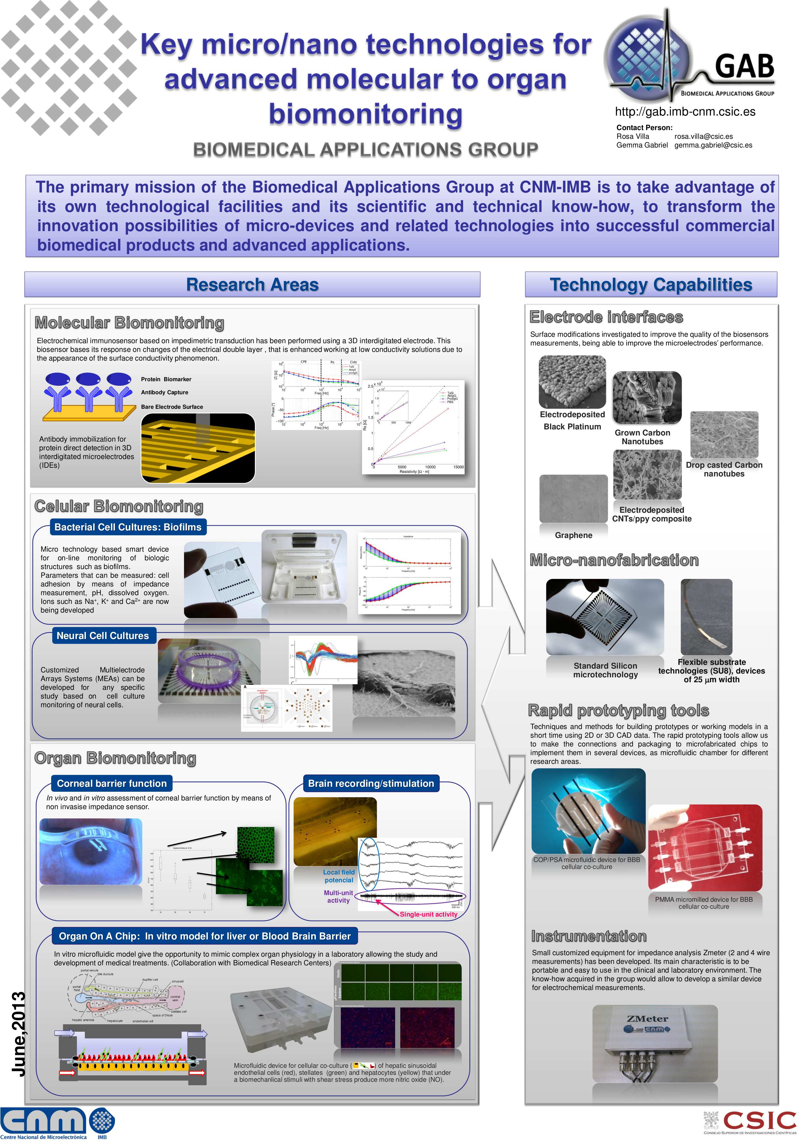
Scientists at the CSIC and at CIBER have developed a device which combines microelectronics and microfluidics to reproduce cell barrier conditions and to monitor them. It has applications on medical essays, to test drugs designed to penetrate cell barriers, such as antitumoral drugs for brain tumors or treatments for neurodegenerative diseases.
Cell cultures are a promising alternative to avoid animal testing. Nevertheless, they have an unresolved issue: they are static cultures, which don’t reproduce the dynamic factors the cells are exposed to in an alive organ or tissue, factors like pressure or fluids surrounding the cells.
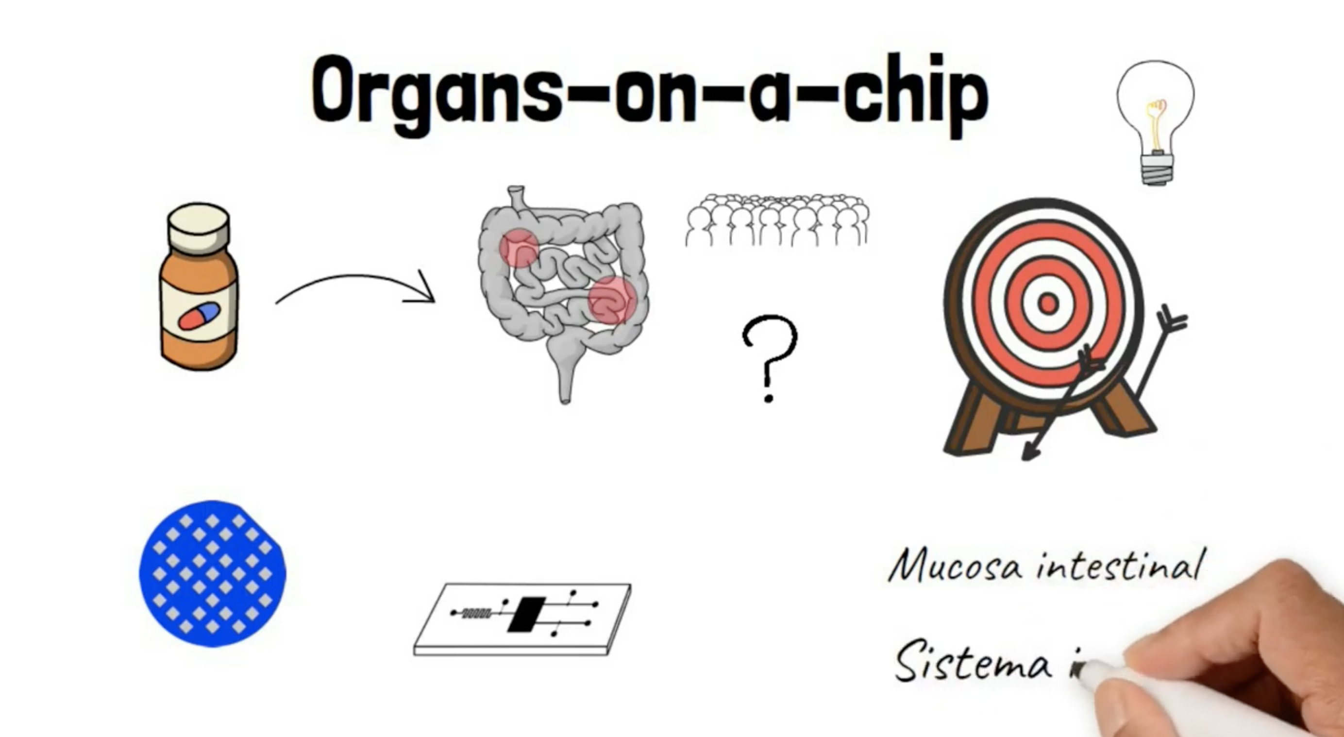
In the last years, microelectronics and microfluidics technologies have evolved enough to reproduce, in the laboratory, the real conditions in which the cells are interacting. It is what has been called “organ-on-a-chip”, which simulates the structure of organs or tissues on a device and enables to study the cells “in vitro” in conditions that are virtually “in vivo”.
Scientists at the Instituto de Microelectrónica de Barcelona (IMB-CNM) of the CSIC, and at biomedical research center CIBER (Centro de Investigación Biomédica en Red) have developed a device which simulates the blood–brain barrier (also called hematoencephalic barrier) with flow conditions, monitoring in real-time its functionality.
“The blood-brain barrier protects the brain and avoids anything to penetrate except for some substances like oxygen, glucose and alcohol. This barrier is also the reason why it’s so difficult to carry antitumoral drugs to the brain”, explains Rosa Villa, CSIC scientist at the IMB-CNM.
Previous studies have tried to reproduce cell barriers on static culture cells. But it was discovered, explains Rosa Villa, that endothelial cells of the barriers become “more permeable” when they are not exposed to the pressure of the constant flow –blood flow- they have in real conditions within the body. Therefore, experimentation with static culture cells can produce false results as the cells don’t behave as they would do inside the body.
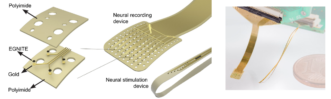
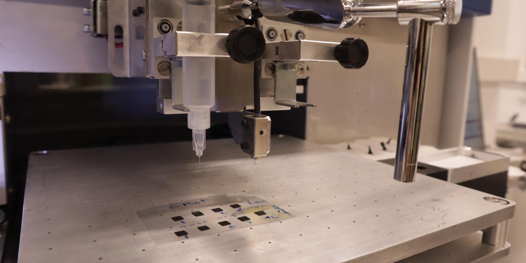
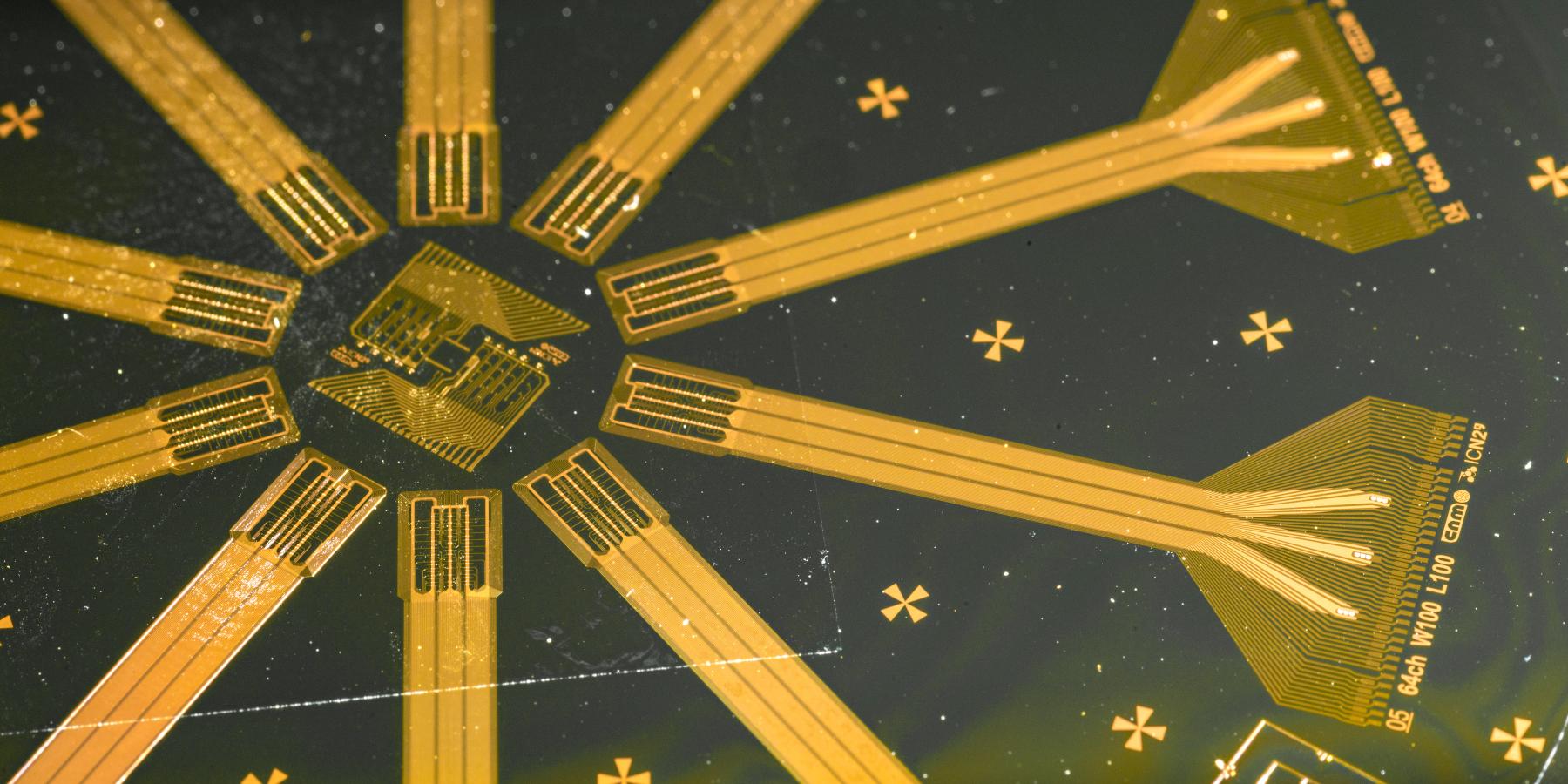
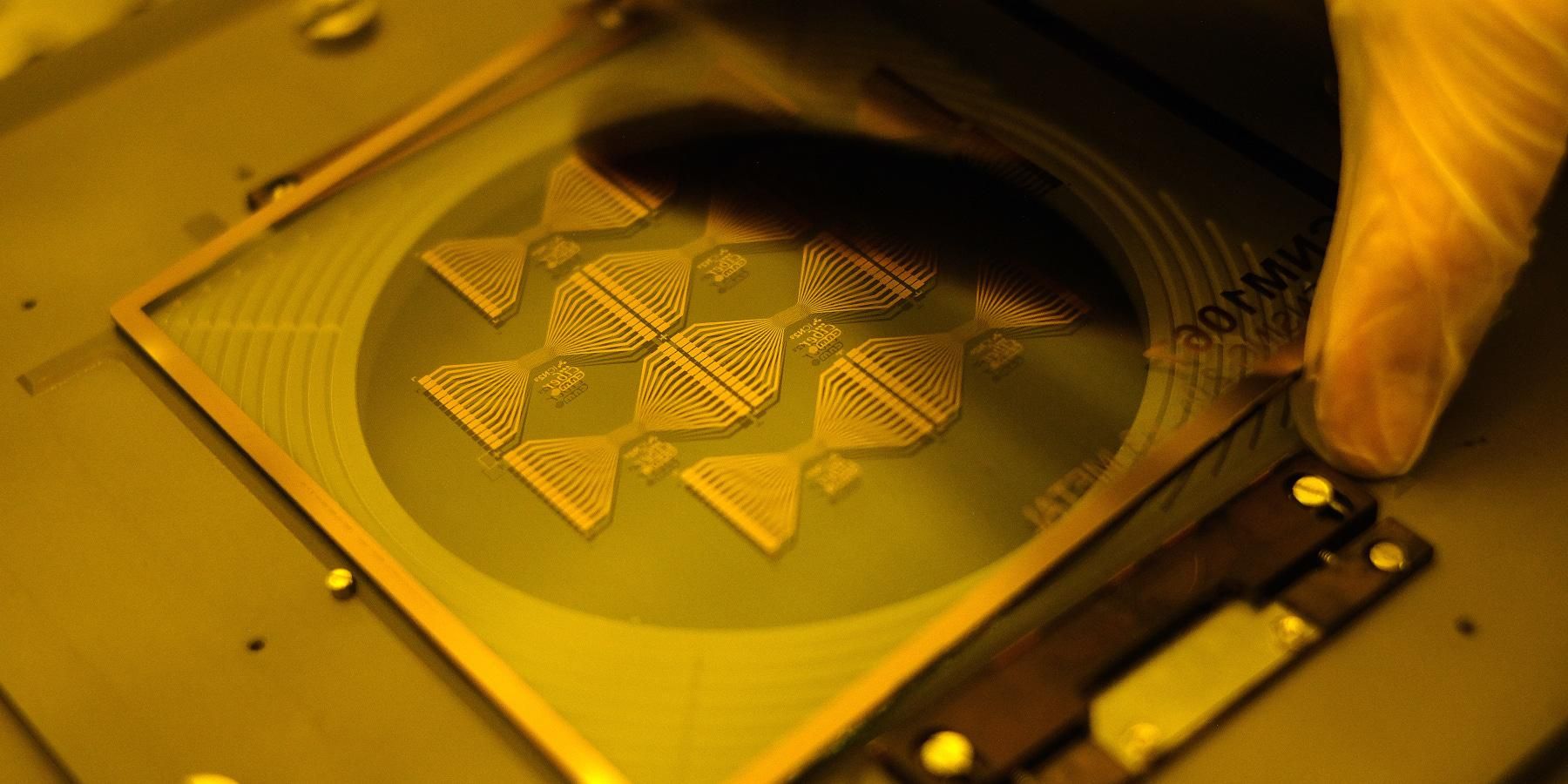
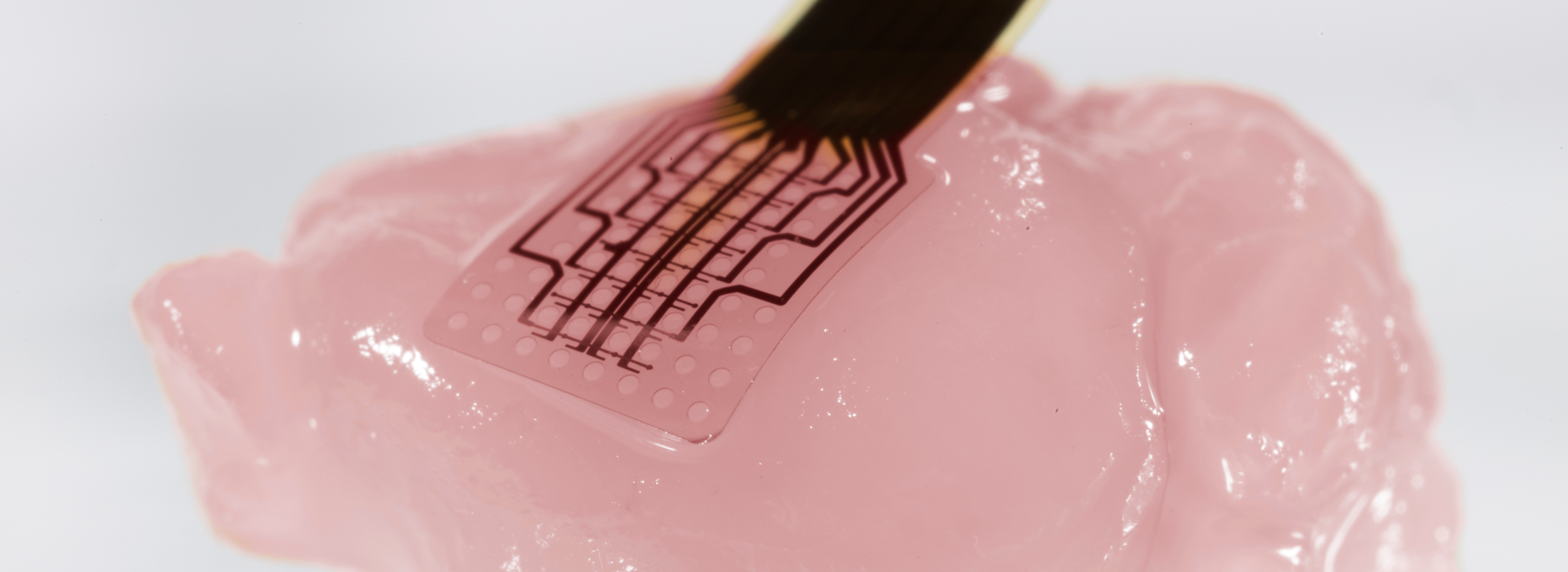
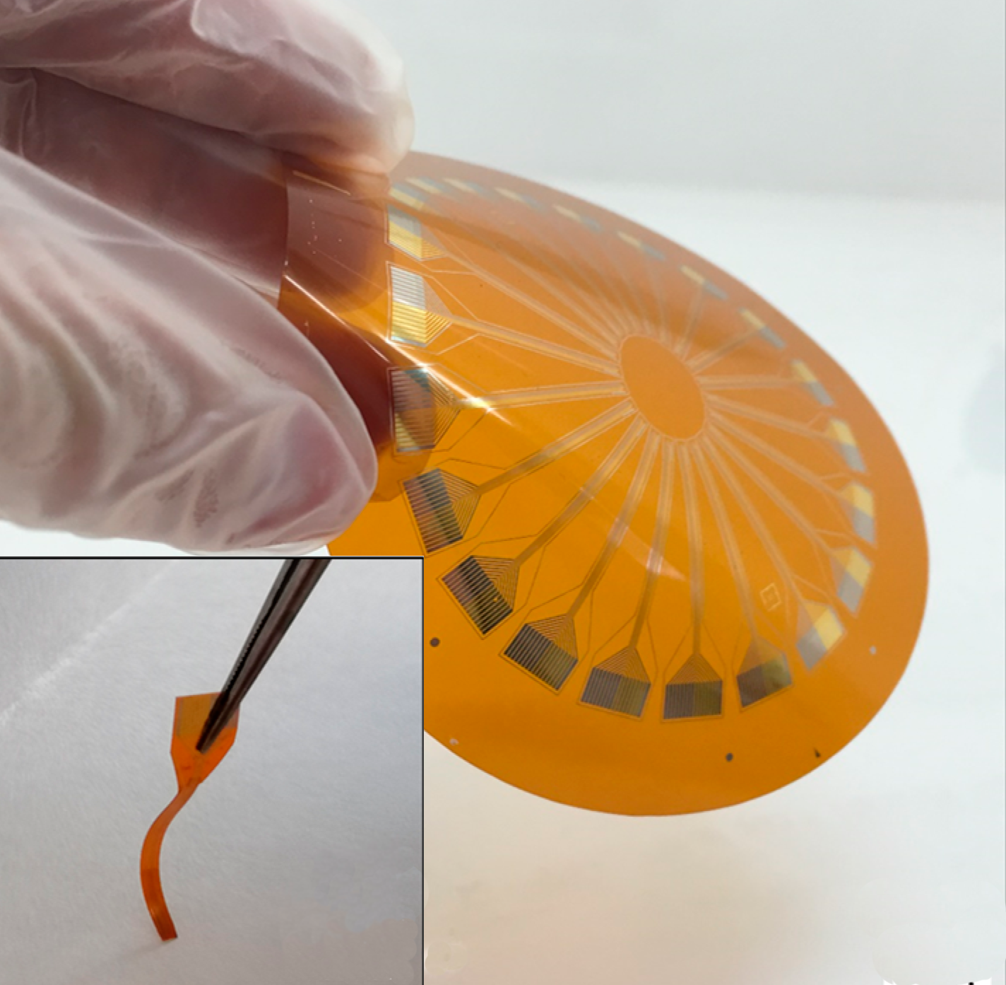
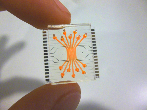
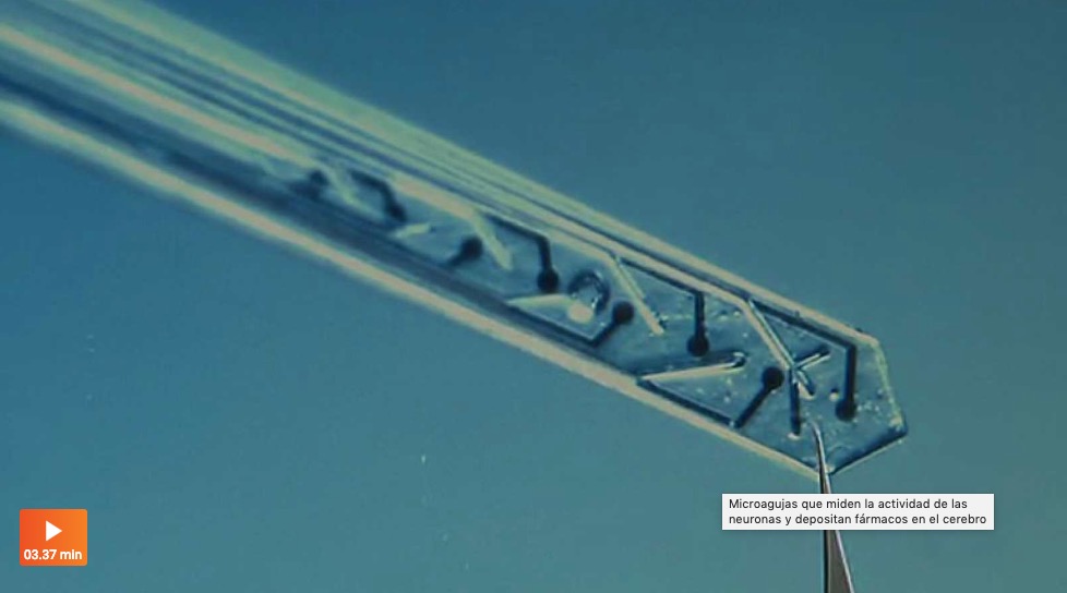
The device is composed of a microfluidic system (the fluid is a serum enriched with nutrients which emulates blood) that reproduces the flow of a cell barrier. It has an electrode system to monitor on real-time the permeability of the barrier through TEER (Transendothelial/Epithelial Electric Resistance) measures. At the same time, it enables visualizing the cell culture. The device has been validated in blood-brain barrier model using animal brain endothelial cells.
It can be scaled to different geometries and applied for experimentation with different cell types. Besides, the microfluidic system is reusable, which reduces the laboratory material expenses. Only the membrane where cell cultures are placed is single-use. The device is scalable, detachable and gives easy access to the inner membrane. And, most important, it is a more reliable tool for essays than static culture cells.
Several laboratories have already asked for the device and have used it in essays. It can be applied for experimenting new drugs that have to penetrate cell barriers, such as treatments for neurodegenerative diseases, or for brain tumors.
16/03/2015